SEMINAR
The State Key Lab of
High Performance Ceramics and Superfine Microstructure
Shanghai Institute of Ceramics, Chinese Academy of Sciences
中 国 科 学 院 上 海 硅 酸 盐 研 究 所 高 性 能 陶 瓷 和 超 微 结 构 国 家 重 点 实 验 室
Progress on Time-resolved Luminescent Probes and Devices
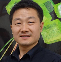
Prof. Dayong Jin
E-mail address: (dayong.jin@uts.edu.au)
Institute for Biomedical Materials and Devices (IBMD), Faculty of Science, University of Technology Sydney, NSW, 2007, Australia
Advanced Cytometry Labs, ARC Centre of Excellence for Nanoscale BioPhotonics (CNBP), Macquarie University, Sydney, NSW, 2109, Australia
时间:2015年12月23日(星期三)上午 9:00
地点:2号楼607会议室(国家重点实验室)
欢迎广大科研人员和研究生参与讨论!
联系人:施剑林(2712)、步文博(2610)
报告人简介:
 Professor JIN, Dayong is an Australian Research Council Future Fellow in photonics, nanotechnology & medical biotechnology, the Chief Investigator of ARC Centre of Excellence for Nanoscale Biophotonics. Since 2008, Prof. Jin established the Advanced Cytometry Labs @ Macquarie University, where he developed a new material library of multi-functional Super Dots? for rapid diagnostics, nanoscale molecular imaging and anti-counterfeiting applications. For his pioneering work in tailoring the multidisciplinary approaches to develop the simple method for detecting hidden, diseased cells, his team has been awarded the 2015 Australian Museum UNSW Eureka Prize for Excellence in Interdisciplinary Scientific Research. In 2015, Prof. Jin accepted an offer to join the University of Technology Sydney as a chair professor to lead its Materials and Technology research strength and grow the UTS Institute of Biomedical Materials & Devices. His goal by 2020 is to develop a more dynamic collaborative research hub which is featured by solution-driven research. http://www.uts.edu.au/about/faculty-science/news/new-collaborative-science-expert-put-ibmd-world-stage.
报告摘要:
Detection, quantification, or localisation of particular cells or molecules – quickly, sensitively and accurately – is fundamental to many areas of modern biomedical research and industry: from understanding sub-cellular processes and biochemical pathways to the early detection of diseases in clinical settings. However, molecules that are altered as a result of a pathological condition are generally present in low abundance, and are difficult to detect. This is particularly true in the early stages of disease development or infection outbreak, posing a “needle-in-a-haystack” problem that demands sensitivity, resolution, and speed.
My talk will showcase our research roadmap from developing bothluminescent materials and photonic sensing techniques to integrated biomedical devices. In 2011, we first developed a low-cost time-gated chopper unit, compatible to any fluorescence microscopes, to realize background-free imaging of rare cells. [1] In 2012, we developed a high speed scanning cytometry for rare-event detection in noisy backgrounds with higher sensitivity than flow cytometry. [2] In 2013, we have overcome the barrier of concentration quenching to address the brightness limitation of upconversion luminescence probes, and created the brightest nanocrystal for single molecule sensing. [3] In 2014, we reported a new lifetime scanning technique [4] anda library of luminescent probes with tunable microsecond lifetimes (t), offering significant advantages in high speed and high throughput sensing of multiple analytes in a single test (multiplexing). [5]
This year, we have madenew progresses in the controlled synthesis of heterogeneous nanocrystals, bio conjugation, and device engineering.We demonstrated a fabrication technique for producing heterogeneous nanoparticles with desirable size, shape,surface, and composition placements with atomic scale precision. [6] This control enables selective epitaxial growth of shells over nanocrystal cores, to form a diverse library of monodisperse 3-D nanoparticles. In parallel, we developed a simple and rapid protein bio-conjugation method based on a one-step ligand exchange using the DNAs as the linker. [7]This method can efficiently pull out the upconversion nanoparticles from organic solvent into an aqueous phase, and the upconversion nanoparticles then become hydrophilic, stable and specific biomolecules recognition. In instrumentation, we engineered an on-the-fly stage scanning method to achieve precise target pinpointing across the whole slide. [8]It can search a coverslip area in just 5.3 minutes, and precisely pinpoint individual targets for precise quantification of luminescence intensities, colours, and lifetimes of single microspheres, crystalline microplates, and colour-barcoded microrods, as well as quantitative suspension array assays of biotinylated-DNA functionalized upconversion nanoparticles.
Our work have paved solid foundations for advanced applications of time-domain biophotonics, thereby solving five key problems, low signal strength, specificity and speed, along with background interference and large-scale multiplexing capacity, that currently prevent rare-event detection, quantification and visualization and are presently limiting the discovery of new biomarkers and development of diagnostic tests.
References
- Jin D, Piper J, Analytical Chemistry 83 (6), 2294–2300 (2011)
- Lu J, Martin J, Lu Y, Zhao J, Yuan J, Ostrowski M, Piper JA, Paulsen IT, Jin D*, Analytical Chemistry, 84 (22), pp 9674–9678 (2012)
- Zhao J, Jin D*, Schartner E, Lu Y, Liu Y, Zhang L, Zvyagin A, Dawes J, Nature Nanotechnology, 8, 729-734, 2013.DOI: 10.1038/nnano.2013.171
- Lu Y, Lu J, Zhao J, Cusido J, Raymo F, Yuan J, Yang S, and Jin D*, Nature Communications, DOI: 10.1038/ncomms4741
- Lu Y, Zhao J, Zhang R, Liu Y, Liu D, Goldys E, Robinson J, Jin D*, Nature Photonics, 8, 32-36, 2014. doi: 10.1038/nphoton.2013.322.
- Liu D, Xu X, Du Y, Qin X, Zhang Y, Ma C, Wen S, Ren W, Jin D*, Nature Communications,DOI: 10.1038/ncomms10254,in press
- Lu J, Chen Y, Liu D, Ren W, Piper J, Paulsen I, Jin D*, Analytical Chemistry, 2015, 87 (20), pp 10406–10413, DOI: 10.1021/acs.analchem.5b02523
- Zheng X, Lu Y, Zhao J, Zhang Y, Leif R, Liu X, Jin D*, Analytical Chemistry, 2015, in press


 当前位置:
当前位置:

